Heart Anatomy
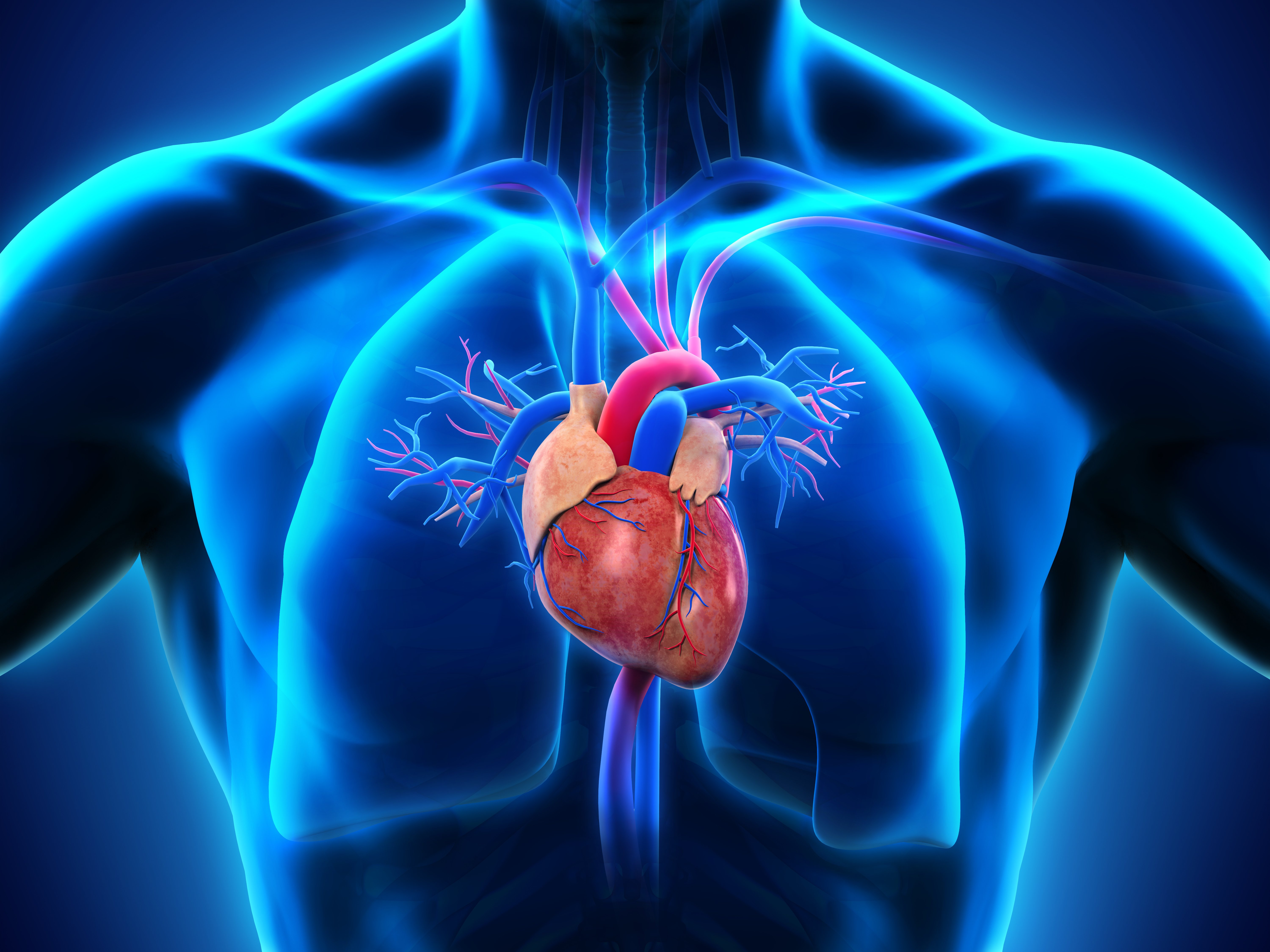
The heart is a muscular organ in most animals.This organ pumps blood through the blood vessels of the circulatory system. The pumped blood carries oxygen and nutrients to the body, while carrying metabolic waste such as carbon dioxide to the lungs. In humans, the heart is approximately the size of a closed fist and is located between the lungs, in the middle compartment of the chest, called.
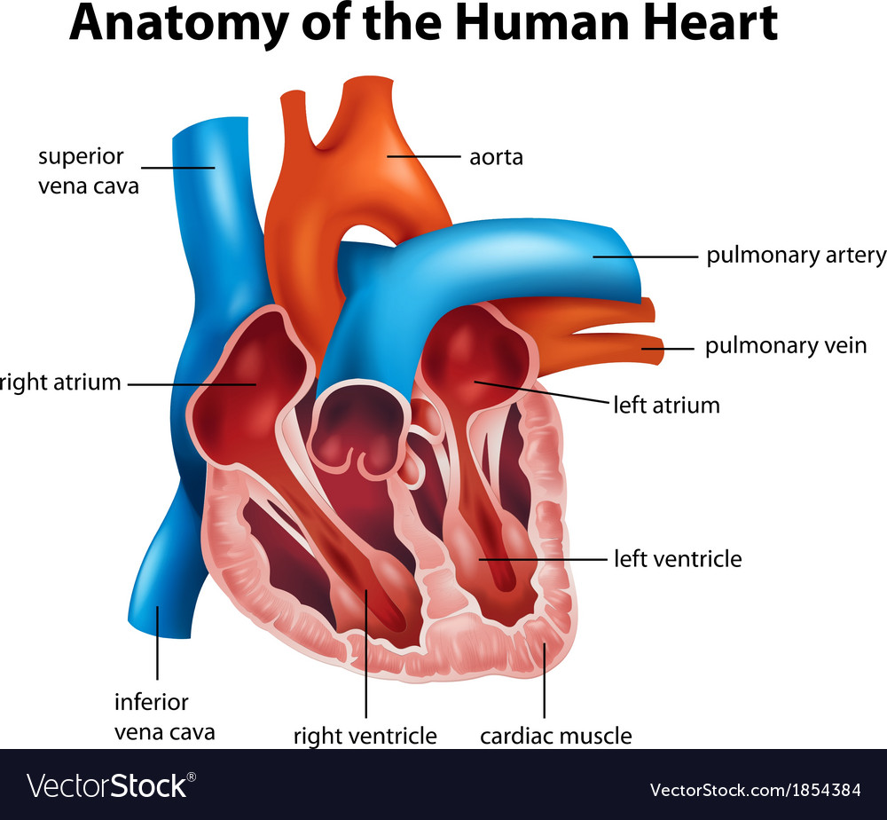
Human heart anatomy Royalty Free Vector Image VectorStock
By: Tim Taylor Last Updated: Jul 30, 2020 2D Interactive NEW 3D Rotate and Zoom Anatomy Explorer Heart Aortic Valve Bundle Branches Chordae Tendineae Interventricular Septum Left Atrium Left Auricle Left Ventricle Mitral Valve Papillary Muscles Pulmonary Valve Purkinje Fibers Right Atrium Right Auricle Right Ventricle Tricuspid Valve
On Heart Kardiohirurgija.rs
Anatomy of the human heart and coronaries: how to view anatomical structures. This tool provides access to an MDCT atlas in the 4 usual planes, allowing the user to interactively discover the heart anatomy. The images are labeled, providing an important medical and anatomical tool. The quiz mode makes it possible to evaluate the user's progress.

Heart Anatomy Stock Illustration Download Image Now iStock
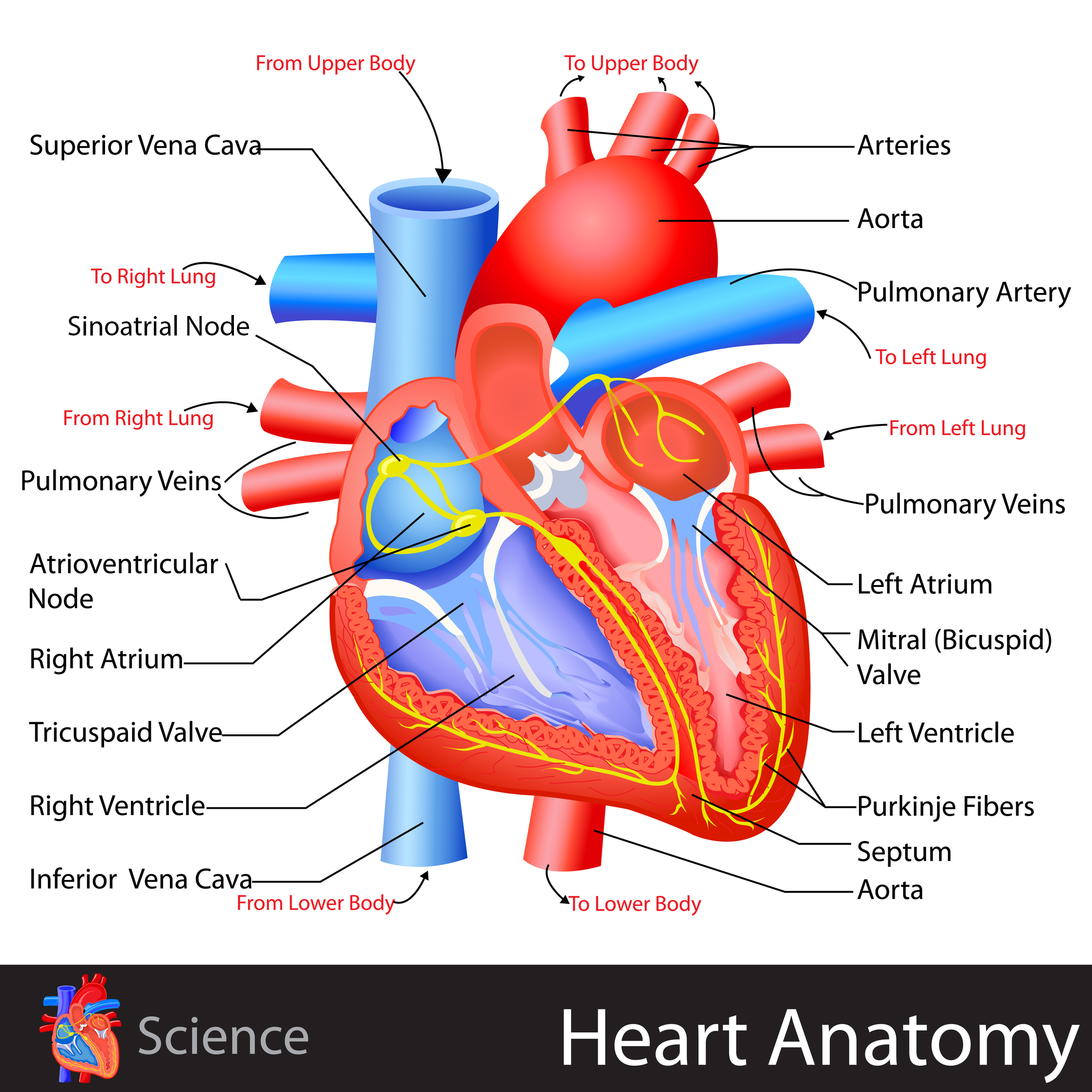
The heart wall consists of three layers: Epicardium: The outer layer of the wall of the heart. Myocardium: The muscular middle layer of the wall of the heart. Endocardium: The inner layer of the heart. Cardiac Conduction Cardiac conduction is the rate at which the heart conducts electrical impulses.
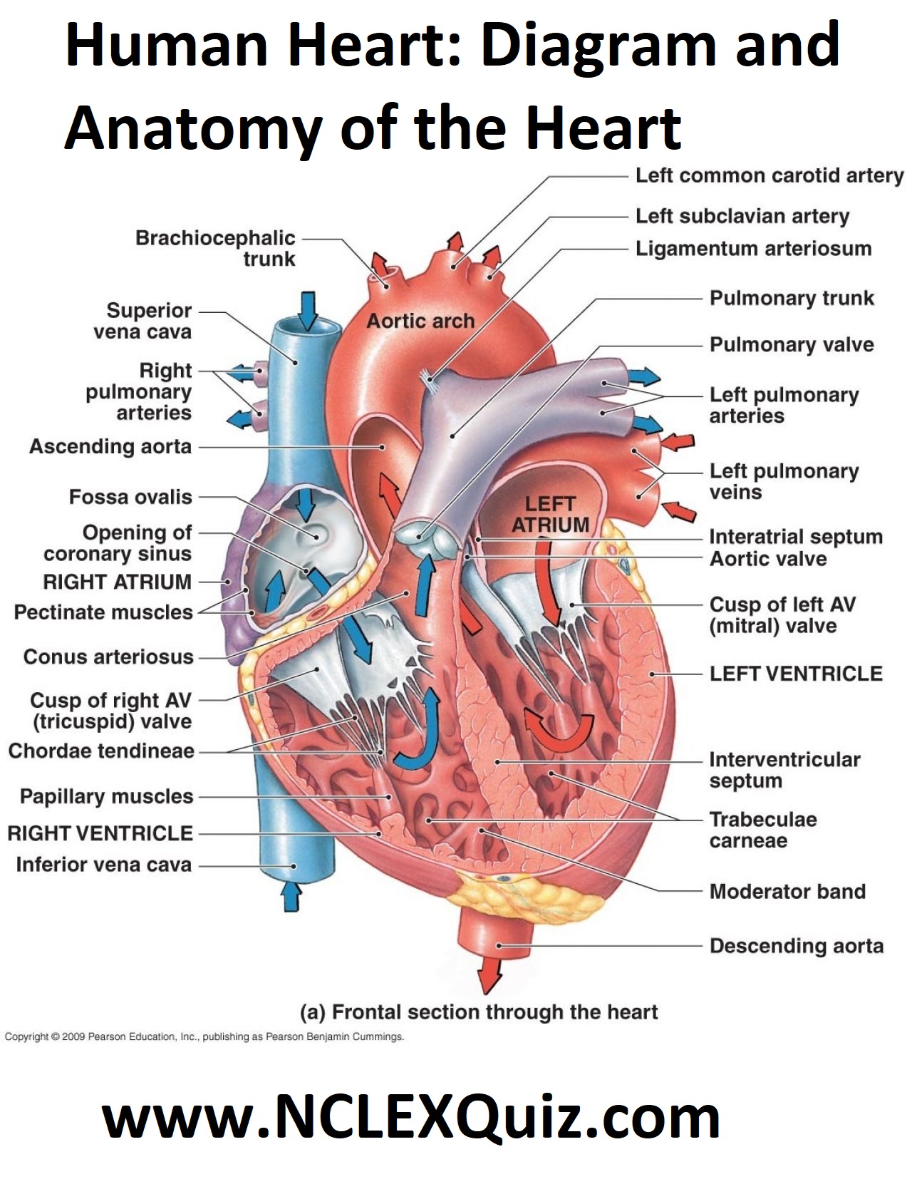
Human Heart Diagram and Anatomy of the Heart StudyPK
It may be a straight tube, as in spiders and annelid worms, or a somewhat more elaborate structure with one or more receiving chambers (atria) and a main pumping chamber (ventricle), as in mollusks. In fishes the heart is a folded tube, with three or four enlarged areas that correspond to the chambers in the mammalian heart.

Human Heart Anatomy Diagram Health Images Reference
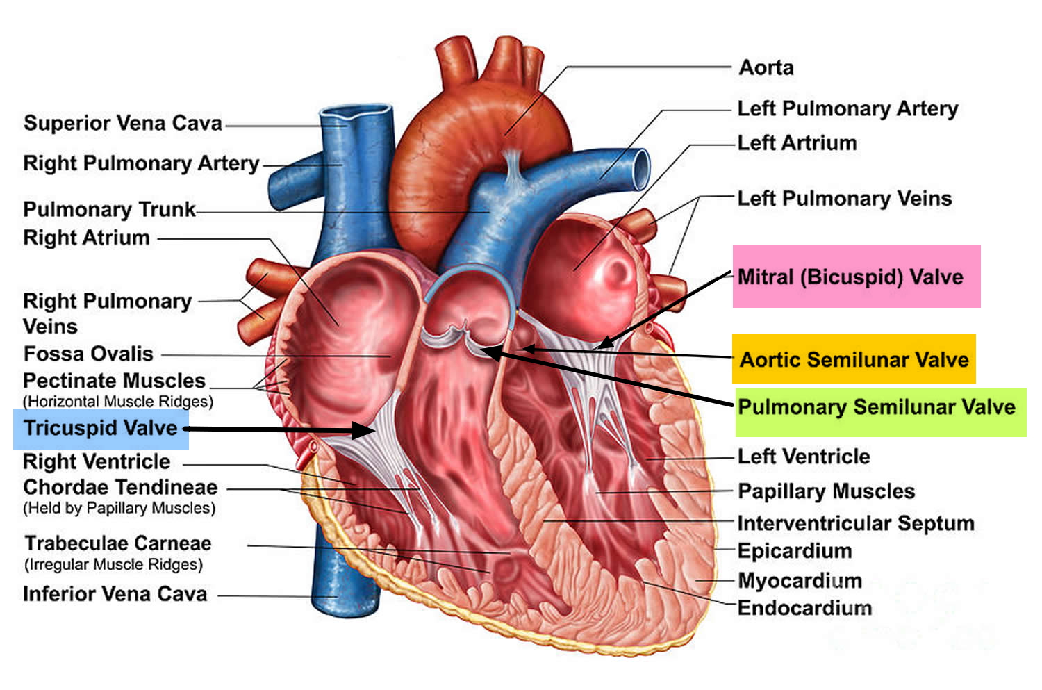
Structure and Function The heart is subdivided by septa into right and left halves, and a constriction subdivides each half of the organ into two cavities, the upper cavity being called the atrium, the lower the ventricle. The heart, therefore, consists of four chambers: right atrium left atrium right ventricle left ventricle [4].
/human-heart-circulatory-system-598167278-5c48d4d2c9e77c0001a577d4.jpg)
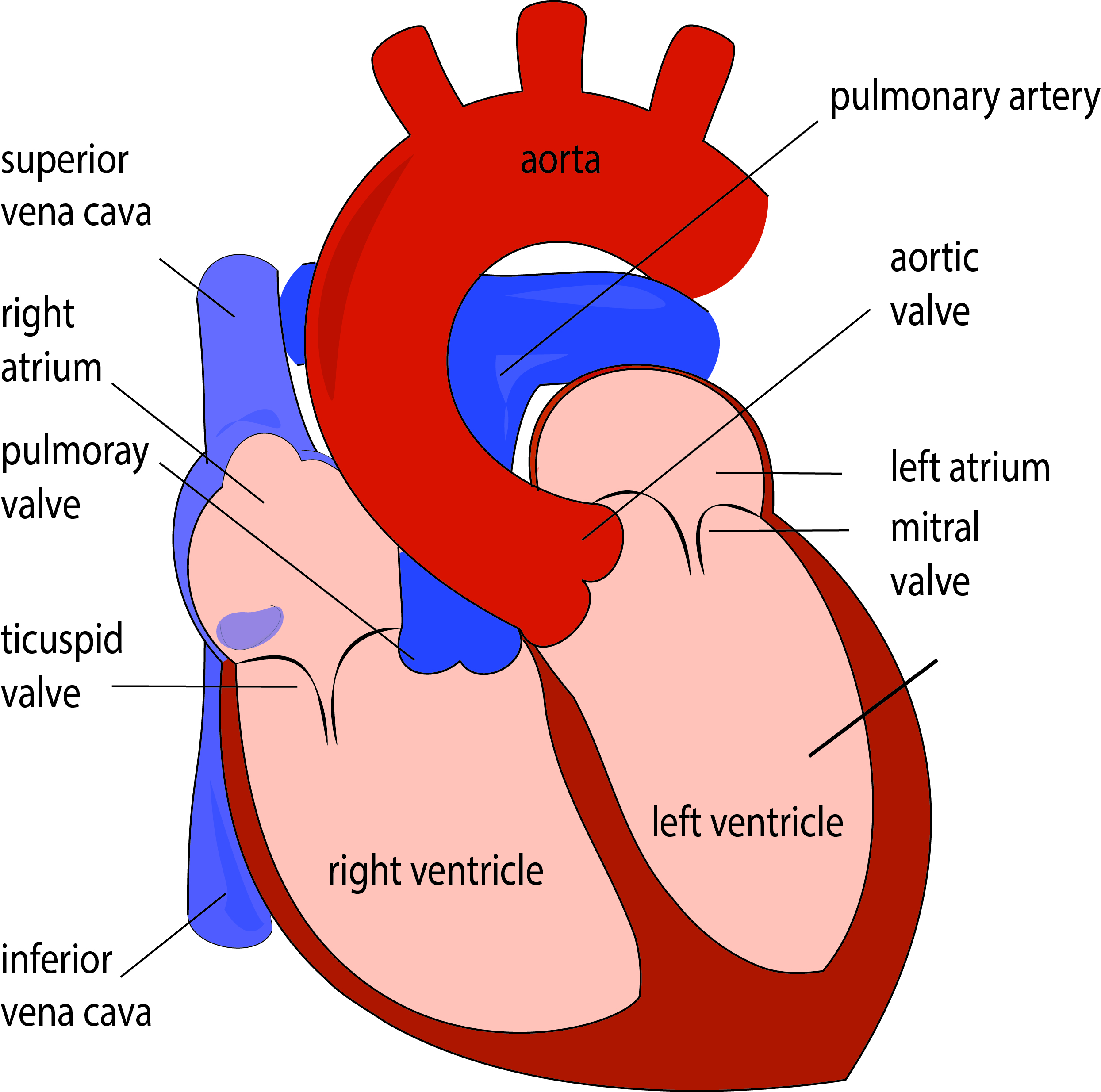
AV and Semilunar Heart Valves
Picture of Heart. The heart is a muscle that pumps blood throughout the body. The heart is located beneath the rib cage slightly to the left of the center of the chest between the lungs and above the diaphragm. A healthy adult heart is approximately the size of a closed fist. The valves and chambers of the heart work together to pump oxygen.
Heart Valves. Function, Purpose and How Many Heart Valves in Your Heart
1. The Heart Wall Is Composed of Three Layers. The muscular wall of the heart has three layers. The outermost layer is the epicardium (or visceral pericardium). The epicardium covers the heart, wraps around the roots of the great blood vessels, and adheres the heart wall to a protective sac. The middle layer is the myocardium.

Cardiovascular Disease
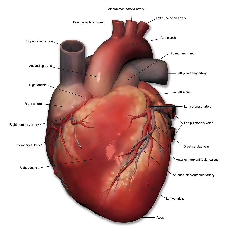
Great vessels of the heart Clinical notes Sources Related articles + Show all Heart anatomy The heart has five surfaces: base (posterior), diaphragmatic (inferior), sternocostal (anterior), and left and right pulmonary surfaces. It also has several margins: right, left, superior, and inferior:

Heart Anatomy chambers, valves and vessels Anatomy & Physiology
AHA/ASA Medical Graphics and Illustrations. Atherosclerosis blockage forming graphic. Download (102.0 kB) Blood clot breaking off in carotid artery. Download (93.3 kB) Blood clot formation in carotid artery. Download (97.5 kB) Blood clot in the brain. Download (95.0 kB)
Anterior View Of Human Heart Anatomy Photograph by Alayna Guza Pixels
The heart is an organ that weighs approximately 350 grams (less than one pound). It's nearly the size of an adult's clenched fist. It's located in the thorax (chest)—between the lungs —and extends downward between the second and fifth intercostal (between the ribs).

Infographic Anatomy of the Human Heart Kids Discover
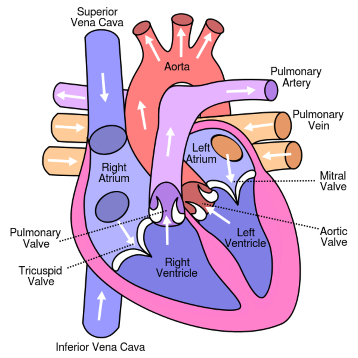
The heart is divided into four chambers: two atria and two ventricles. Blood is transported through the body via a complex network of veins and arteries. The average human heart weighs between.

Anatomy of the human heart Royalty Free Vector Image
Summary. The heart is a complex organ that pumps blood throughout the body. It sits in the chest, slightly left of center, behind the breastbone, and between the lungs. A heart that is not healthy.
Learn About the Heart and Circulatory System for Kids HubPages
Cardiovascular System Anatomy The Heart. The heart is a muscular pumping organ located medial to the lungs along the body's midline in the thoracic region. The bottom tip of the heart, known as its apex, is turned to the left, so that about 2/3 of the heart is located on the body's left side with the other 1/3 on right.
Human Heart Anatomy APDA
What is the heart? The heart is a fist-sized organ that pumps blood throughout your body. It's the primary organ of your circulatory system. Your heart contains four main sections (chambers) made of muscle and powered by electrical impulses. Your brain and nervous system direct your heart's function. Advertisement

Heart Valves. Function, Purpose and How Many Heart Valves in Your Heart
Anatomy of the interior of the heart. This image shows the four chambers of the heart and the direction that blood flows through the heart. Oxygen-poor blood, shown in blue-purple, flows into the heart and is pumped out to the lungs. Then oxygen-rich blood, shown in red, is pumped out to the rest of the body, with the help of the heart valves.